|
Macular Degeneration The Gradual Loss of the Fine Details of Life
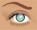
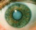
Macular degeneration (MD) — also known as age-related macular degeneration (AMD) — is the gradual loss of central vision (including fine detail vision) due to damage to and subsequent breakdown of the macula, the small central portion of the eye. The macula is the yellow-colored area of The Retina; it is packed with cone receptors, responsible for color detection and fine detail vision. There are two types of MD: dry (or atrophic) MD and wet (or exudative) MD. Dry MD is the most common form, accounting for about 85% of all cases in North America. It causes a gradual deterioration of vision. There is no blood, fluid or protein leakage with this type, leading to the term “dry” MD. Due to a buildup of waste products (protein deposits) called drusen, retinal pigment epithelial (RPE) cells above the drusen are lost, resulting in the death of the photoreceptors that depended on the RPE cells for nourishment. Eyesight Loss in that area is the result. Large, thick drusen also increase the risk of developing wet MD. Wet MD is less common. Wet MD occurs when blood vessels from the choroid (the layer that the Bruch’s membrane and RPE cells rest on) grow through the Bruch’s membrane and under the RPE. These blood vessels do not have tight junctions, and tend to leak, letting blood and other fluids escape. Once the vessels grow through Bruch’s membrane, they can balloon rapidly, leading to damage to the RPE and the photoreceptors. The result is rapid vision loss.
Causes
MD is caused by a variety of factors, most related to damage by Free Radicals. Visible light, particularly the blue wavelengths, can damage the macula. The yellow color of the macula is from the yellow pigments present there. These pigments absorb the blue wavelengths, so when pigment levels diminish, more radiation can damage the cells of the Retina. Poor blood flow can result in oxygen or nutrient starvation, which then triggers chemical signals that cause abnormal blood vessel growth in the choroid. Elderly people have been shown to have less than two-thirds of the blood flow in the back of the eye that younger people have. Reduced blood flow can be the result of hardened arteries or weakened capillaries.
Symptoms
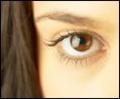
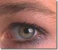
Symptoms of MD include difficulty reading and seeing signs, letters missing from the middle of a page of text, Blurred Vision, distortion, and Loss of Central Vision. Blurred Vision is the most common symptom, lasts for and intensifies over the years, and becomes more difficult to deal with. Early on, stronger glasses and better lighting can overcome the reading problem, but eventually even low-vision aids such as magnifiers can’t help. You may notice distortions (metamorphopsia) when you look at vertical linear objects, such as utility or light poles, or vertical blinds. The object will look wavy or crooked. This is caused by blood vessels growing under the retina, changing its surface from flat to uneven. This results in cone photoreceptors aimed in various directions. This symptom indicates development of wet MD. An Eye Care Exam should occur within 24 to 48 hours. Central vision loss is a later stage of development. One eye is usually affected first, with the other eye losing vision over the next few years. Peripheral vision is not affected, and total blindness rarely occurs. Check out this Vision Simulator if you would like to see what vision deterioration due to MD looks like. The simulator will open in a new window. Close the window to return to this page.
Eye Care ExamTesting
If you want to test yourself for wet MD, use the Amsler Grid, a ruled grid with a dot in the center. Try taping it to your fridge or above your computer monitor, where you can test your vision for MD regularly. Cover one eye, then focus on the dot. Lines appearing wavy or distorted, or dark spots or missing areas apparent in the grid, indicate that wet MD may be developing. If you notice any of these symptoms, call your Eye Care Professional or Eye Care Specialist immediately, as prompt treatment may preserve your reading vision. Eye doctors also use a slit lamp exam to look for drusen and fluids. The appearance of fluids requires the use of a fluorescein angiogram (FA). The FA shows where the abnormal blood vessels are. The doctor can use the FA as a road map for laser treatment of macular degeneration.
Risk Factors Risk factors for macular degeneration include:
Treatment and Cure There is no cure for macular degeneration, and the only treatment is for wet MD. This treatment only stops further deterioration; it cannot restore lost vision. Laser treatment involves pulsing high-energy light through the Cornea and lens to the retina, where the abnormal blood vessels are “cooked.” The coagulation of blood in the cooked vessels stops the blood flow and prevents further growth. However, the parts of the retina around the blood vessels are permanently damaged and become scar tissue. Followup visits every few weeks are necessary, to check for growth of new abnormal blood vessels. Any new growth will require further laser therapy. A new dry MD therapy, called Reo Therapy, filters certain proteins from the blood and can help with absorption of soft drusen, stabilizing and possibly improving vision. However, this treatment is experimental, very expensive, and does not work with hard drusen. Since most macular degeneration damage is caused by light and free radicals, wearing a good pair of Sunglasses that screen out at least 88% of blue light wavelengths, and ensuring proper nutrition, will go a long way towards reducing central vision loss. Nutritional Help to Prevent or Halt Macular Degeneration
There are several products specifically developed to help slow the progression of macular degeneration, or even prevent it if taken before damage begins. Learn more about these macular degeneraion
products for eye health
here.
For more information check out
Macular Degeneration Books

Subscribe to EyeSight Vision Care! , our monthly newsletter with in depth information to help you keep up to date on how to Protect Your Eyesight with a free bonus. Fill out the form below. You'll then receive an email asking you to confirm that you subscribed. You'll always have the option to unsubscribe at the click of your mouse.
|


More Information