|
Eye Anatomy How the Eyes Work
Eye anatomy think of the eye as a hollow ball, like a tennis ball, with a diameter about the size of a quarter. Most of what makes your sight happen occurs on the inside of your eye. There are three layers in the eye anatomy, what ophthalmologists call tunics. The first layer is the Cornea and sclera. The second layer is the vascular tunic called the uvea, which includes the iris (which surrounds the pupil), the ciliary body, and the choroid. The third layer is The Retina, which rests on the inside surface of the back of the eyeball.
The Cornea and Sclera
The transparent cornea protects your eye anatomy from damage, helps maintain its shape, and, to some extent, bends light toward the lens. The cornea is surrounded by the opaque sclera, the white of the eye anatomy, which surrounds the entire eyeball and also helps maintain the eye’s shape. The cornea and sclera are both made of collagen, which gives this layer its toughness and ability to maintain the shape of the eye.
The Iris, Ciliary Body, and Choroid
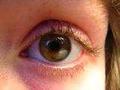
The iris, the colored part of the eye, surrounds the pupil. The pupil is, in a sense, nothing. The pupil is the hole at the front of the eyeball where light enters the eye anatomy. The size of this hole is regulated by the iris. The iris shrinks the size of the pupil in bright light (to reduce damage caused by too much radiation), and dilates it in dim or dark conditions (so that as much light as possible enters the eye). The ciliary body is the set of muscles and ligaments that control the shape of the lens so that light from both nearby and distant objects can be focused onto the Retina. The ciliary body also produces the aqueous humor, a thin fluid that fills the space between the cornea and the lens. The aqueous humor provides glucose, proteins and oxygen to the cornea, iris and lens. The choroid contains the small arteries that supply oxygen and nutrients to the front of the eye anatomy. It is also made up of the small veins that remove the waste products from this area. The choroid rests on the inside wall of the eyeball.
The Retina
The retina is made up of several parts. The entire retinal layer rests on the choroid, and is separated from it by Bruch’s membrane, a thin film of collagen. Sitting on top of this membrane is the retinal pigment epithelium (RPE). The RPE is a layer of cells that nourish the photoreceptors. The RPE acts in much the same way as the soil in a garden nourishes the plants that grow in it. The photoreceptors are the rod-shaped and cone-shaped cells that convert the light entering your eye into electrical signals that are passed on to the brain. There are about six million cones and 120 million rods. The cones allow detailed vision, central vision, and colors (in brightly lit circumstances). The rods are responsible for all Night Vision and peripheral vision. Photoreceptors wear out, and are constantly replaced with the help of the RPE. They contain a protein called rhodopsin (also called retinal purple and visual purple), which contains Vitamin A. They also contain high concentrations of polyunsaturated fats. On the tops of the photoreceptors are nerve cells and fiber layers. These cells transmit the signals created by the photoreceptors to the optic nerve. Retinal blood vessels in the nerve cell layers provide these cells with nutrients. Retinal glial cells provide structural support inside the retina.
The Two Chambers
There are two chambers, or compartments, inside the eye. The anterior (meaning front) chamber is the space between the cornea and the lens/ciliary body. It's filled with the aqueous humor, a fluid that provides the nutrients to the front parts of the eye, and maintains the internal pressure of the eye. (The lens has no nerves or blood vessels, and so is completely dependent on the aqueous humor for its nourishment and removal of waste products.) The Vitreous cavity takes up the middle and back of the eye. The vitreous humor, which fills the remaining 80% of the hollow part of the eye, is a clear substance with the texture of an egg white. It is made up of collagen, a protein.
On the Outside
On the outside of the eye are the extraocular muscles, a set of six muscles that control the movement of the eye up and down, side to side, and from the top left to the bottom right and vice versa. The eyelid and eyelashes protect the eye from foreign bodies and too bright light. The tear duct lubricates the outer surface of the eye so that the cornea is not damaged. The eyeball rests in the orbital socket. This socket is lined with fatty tissues, which protect the eyeball from trauma. Also on the outside is the optic nerve, which carries the electrical signals to the visual cortex of the brain.

Subscribe to EyeSight Vision Care! , our monthly newsletter with in depth information to help you keep up to date on how to Protect Your Eyesight with a free bonus. Fill out the form below. You'll then receive an email asking you to confirm that you subscribed. You'll always have the option to unsubscribe at the click of your mouse.
eye anatomy - protect your eyesight
|


More Information